Journal of Regional Section of Serbian Medical Association in Zajecar

Year 2025     Vol 50     No 1-2
     
      [ Contents ] [ INDEX ] >>> ]      
       

Page 6


Original paper

Rotational thromboelastometry guided blood component administration

Radovan Dinić (1), Almira Šabani (1), Jasmina Grujić (2)

1) HOSPITAL BLOOD BANK, DEPARTMENT OF PRE-TRANSFUSION TESTING, EMERGENCY CENTER OF THE UNIVERSITY CLINICAL CENTER OF SERBIA, BELGRADE; 2) DEPARTMENT OF TRANSFUSIOLOGY, FACULTY OF MEDICINE, UNIVERSITY OF NOVI SAD, NOVI SAD


  Download in pdf format   Abstract: Introduction: Rotational thromboelastometry is a diagnostic tool extensively utilized for patients at high risk of bleeding, particularly those experiencing trauma or undergoing significant surgical procedures. This test is notable for providing rapid results, facilitating timely decisions regarding the application of individualized transfusion therapies, thereby optimizing the rational use of blood components. Aim: The objective of this study is to assess the utilization of blood components in the transfusion treatment of patients at the Emergency Center of the University Clinical Center of Serbia, based on the outcomes of rotational thromboelastometry. The analysis aims to evaluate the frequency of these interventions in the studied population and to highlight the advantages of this treatment modality.
Material and methods: This retrospective study analyzed trauma patients who underwent rotational thromboelastometry at the Hospital Blood Bank, Department of Pre-transfusion Testing, Emergency Center of the University Clinical Center of Serbia, from January 1st 2023, to December 31st 2023. Data regarding the results of the thromboelastometry tests and subsequent blood component therapies were extracted from the Service's protocols. Results: During the study period, 776 patients were evaluated by means ofrotational thromboelastometry, of which 358 (46.13%) required blood component therapy. Cryoprecipitate was administered to 48 (13.40%) patients, platelet concentrate to 69 (19.27%), and a combination of cryoprecipitate and platelets to 61 (17.03%) patients. Additional therapeutic approaches included the administration of platelets, desmopressin, and tranexamic acid, while fresh frozen plasma was utilized the least, in only 17 (4.74%) patients. Conclusion:The analysis of blood components used for therapeutic purposes in relation to the rotational thromboelastometry demonstrated a wide range of therapeutic modalities in the treatmentof patients. This testing method facilitates individualized therapy with blood components, subsequently diminishing the needfor transfusions, enhancingallows for better diagnostic precision, and reduces costs associated with long-term managementof patients.
Key words: rotational thromboelastometry, therapy, trauma, transfusion, bleeding


INTRODUCTION

Rotational thromboelastometry (ROTEM) is a test whichis an integral part of "Point-of-Care" testing. It is characterized by being performed within a short time frame after the patient's sample is taken, allowing timely decisions regarding the transfusion management of the patient. The results are obtained quickly, which enables a rapid modification of therapy if needed [1]. ROTEM testing is being increasingly integrated into the routine diagnostic algorithm and management of bleeding in patients at high risk of bleeding, such as trauma patients. At the Emergency Center of the University Clinical Center of Serbia, the majority of patients are those admitted urgently as a result of trauma. A rapid clinical assessment of the injured patient's condition allows the prediction of trauma-induced coagulopathy (TIC) and the need to activate the massive transfusion protocol [2]. For these purposes, multiple ROTEM tests can be used to distinguish between mechanical and hemostatic bleeding, identify disorders in various stages of the hemostatic process, thereby enabling targeted and rational use of blood and blood components. ROTEM is a functional test that graphically displays the formation and breakdown of a clot, allowing monitoring of the progression or resolution of coagulopathy following a trauma [3].

ROTEM screening tests
The basic screening tests are the EXTEM and INTEM tests. These screening tests provide general information about the status of the hemostasis system. The EXTEM test is sensitive to the deficiency of coagulation factors in the extrinsic pathway, while the INTEM test is sensitive to the deficiency of coagulation factors in the intrinsic pathway, as well as to the anticoagulant effects of heparin and thrombin inhibitors. Both tests are sensitive to platelet participation in clot firmness, fibrinogen levels, fibrin polymerization, factor XIII deficiency, and hyperfibrinolysis [4,5].

Additional ROTEM tests
The analysis of the hemostasis system is expanded by performing additional tests: FIBTEM, APTEM and HEPTEM.
The FIBTEM test is an EXTEM test for the fibrin component of the clot. FIBTEM eliminates the contribution of platelets in clot formation and allows detection of fibrinogen deficiency or fibrin polymerization disorders.
The APTEM test is based on the EXTEM test and allows detection of fulminant hyperfibrinolysis. This test helps identify the need for administering antifibrinolytic therapy. It enables the assessment of whether antifibrinolytic therapy alone normalizes coagulation or if additional measures are required (e.g., administration of fibrinogen or platelets).
The HEPTEM test is an INTEM test that allows identification of hemostasis deficiencies even in the presence of heparin and represents an INTEM test without interference from heparin or heparin anticoagulants [5,6].

The parameters of ROTEM analysis
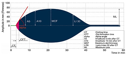
The primary result is a reaction curve in the form of a thromboelastogram, which describes the dynamics of blood clot formation, its size, firmness, and elasticity throughout all the phases of the coagulation process. In routine clinical practice, the following thromboelastogram parameters are analyzed [6].

  1. 1. Clotting Time (CT)
    CT reflects the time from the activation of coagulation to the initial formation of the blood clot, i.e., until the clot reaches a firmness of 2 mm. A prolonged CT is theresult of coagulation factor deficiency, hyperfibrinolysis, hypofibrinogenemia, and the presence of heparin. A shortened CT is a result of hypercoagulability.
  2. 2. Clot Formation Time (CFT)
    CFT reflects the time required to achieve firmness of a 20 mmclot. CFT reflectsthe initial polymerization of fibrin, specifically the interaction between fibrinogen and platelets.
  3. α- Angle
    The alpha angle represents the angle between the horizontal baseline and the tangent that touches the coagulation curve at the point where the firmness of the blood clot of2 mm is achieved. It reflects the quantitative and qualitative relation between fibrinogen and platelets.
  4. Maximum Clot Firmness (MCF)
    MCF represents the maximum amplitude achieved during testing and indicates the stability of the fibrin clot. In the EXTEM and INTEM tests, MCF reflects the levels of fibrinogen and platelets, while in the FIBTEM test it indicates the concentration and functionality of fibrinogen.
  5. Maximum Lysis (ML)
    ML represents the maximum fibrinolytic activity observed during the analysis and indicates the percentage of blood clot lysis.

AIM

The aim of this study is to assess the utilization of blood components in the transfusion treatment of trauma patients at the Emergency Center of the University Clinical Center of Serbia, based on the outcomes of rotational thromboelastometry. The analysis aims to evaluate the frequency of these interventions in the studied population and to highlight the advantages of this type of treatment for patients.

MATERIALS AND METHODS

This retrospective study analyzed trauma patients who underwent ROTEM testing in the Hospital Blood Bank, Department of Pre-Transfusion Testing, at the Emergency Center of the University Clinical Center of Serbia, from January 1st 2023, to December 31st 2023. Based on the obtained results, therapy was administered to each patient. Testing on the ROTEM analyzer was conducted using a tube containing 3.2% sodium citrate as an anticoagulant (Vacutainer Brand, Belliver Industrial Estate, Plymouth, UK, 4.5 ml, 9 NC 0.129 M). A volume of 300 µL of resuspended blood was taken from the tube for each of the two tests (EXTEM and FIBTEM) using a pipette with predefined aspiration and expiration time intervals, it was placed into the "cup & pin" chamber where specific activators for each test were added. Upon activation of each test, the results were monitored on the ROTEM analyzer screen (Figure 1), and once the values were obtained, the result was printed, described, and distributed along with the proposed therapy to the departments that had requested the procedure.

Figure 1. Illustration of the ROTEM graphical record showing all phases of hemostasis.
Sourced from: 10.1515/acm-2015-0006.

Table 1 presents the ROTEM reference values,on the basis of which the therapy was administered to patients.

Table 1. ROTEM parameters, normal values

RESULTS

During the observation period, ROTEM testing was performed on 776 patients. Based on the obtained results, 358 of them (46.13%) required therapy with blood components, while 418 (53.87%) did not need such therapy (Table 2).

Table 2. Need for component therapy based on ROTEM parameter values

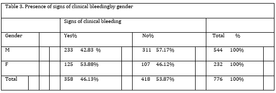
The occurrence of clinical bleeding in patients by gender is shown in Table 3. There is a statistically significant difference in the presence of clinical bleeding related to gender(Χ² = 7.989, p=0.005). Among female patients, 53.88% exhibit clinical bleeding, while 43.83% of male patients show signs of bleeding.

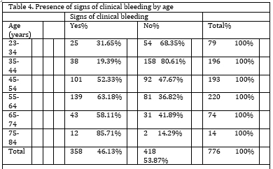
The occurrence of clinical bleeding in patients by age is shown in Table 4. There is a statistically significant difference in the presence of signsofbleeding based on the results of the Χ² test (Χ² = 104.902, p<0.001). Bleeding was most prevalent in the age group of 75 to 84 (85.71%) and least prevalent in the age group of 35 to 44 (19.39%).

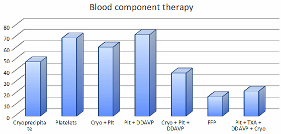
The distribution of blood component usage among patients is shown in Figure 2. Observing the distribution of blood components, we can see that cryoprecipitate was administered to 48 patients (13.40%), platelets to 69 patients (19.27%), and cryoprecipitate and platelets simultaneously to 61 patients (17.03%). The administration of platelets and desmopressin (DDAVP) was necessary for 72 patients (20.11%). Fresh frozen plasma (FFP) was used in only 17 patients (4.74%). A combination of platelets, tranexamic acid (TXA), DDAVP, and cryoprecipitate was administered to 22 patients (6.14%), while cryoprecipitate, platelets, and DDAVP were given to 38 patients (10.6%).

Figure 2. Administered Blood Component Therapy

In 31 patients (8.65%), a tendency towards hypercoagulability was confirmed, and in 11 of these patients (35.48%), the administration of Kybernin P 500 was recommended as a substitution for antithrombin deficiency.

DISCUSSION

The aim of this study was to assess the impact of ROTEM-guided transfusions on the use of blood components in traumapatients. ROTEM-guided therapy with blood components is associated with improved patient outcomes, particularly as it also reduces the need for allogeneic blood transfusions. Our study had a significantly higher proportion of male patients (70.1%) compared to females. Gender differences among trauma patients have been well documented in various studies, with a higher prevalence of males. Specifically, men are more frequently represented as polytrauma patients, often due to risky behaviors that include alcohol consumption and driving under the influence of alcohol [1]. Additionally, most causes for multiple injuries are indeed the result of traffic accidents and work-related injuries, which often occur in younger male populations who are more mobile and engaged in physically demanding jobs [1,2]. Moreover, traumas also occur among the elderly because of falls and fractures [1,3].
Studies have shown that ROTEM can significantly reduce transfusion requirements in cardiac surgery patients, trauma cases, and liver transplantation [7,8]. Görlinger et al. found that the use of ROTEM in cardiac surgery patients decreases the need for red blood cells, FFP and platelets, thereby reducing the risks of complications associated with blood transfusions [9].
The results of our study show that ROTEM-guided transfusion primarily led to increased use of fibrinogen in the form of cryoprecipitate, either as the sole component administered in 13.40% of patients or in combination with other components. Research indicates that ROTEM identifies functional fibrinogen deficiency more reliably than standard hemostasis tests. Several studies have demonstrated that patients with acquired fibrinogen deficiency have increased morbidity and mortality during trauma or procedures that may cause bleeding [10,11]. Schochl et al. demonstrated that ROTEM-guided therapy enables rapid correction of hypofibrinogenemia in severely traumatized patients, reducing the need for large FFP transfusion volumes and associated risks [12]. In cases of trauma-induced coagulopathy (TIC), acquired hypofibrinogenemia is the earliest sign of coagulopathy [9,12]. Fibrinogen levels decrease due to blood loss and hemodilution from the use of crystalloids. A decline in fibrinogen can be observed within the first hour of bleedingas the liver is unable to compensate for such a rapid loss in a short period. [13]. Rapid identification of hypofibrinogenemia is crucial for the effective treatment of coagulopathy.
ROTEM also allows the assessment of platelet deficiency and guides to targeted platelet therapy. The study by Collins et al. demonstrated that ROTEM can identify platelet deficiency in patients with massive bleeding, enabling adequate and timely platelet therapy, thereby reducing the risks of thrombocytopenia and associated complications [14]. Thrombocytopenia is one of the factors predicting the risk of death in trauma patients [9,15]. By using ROTEM, the need for platelet transfusion can be determined shortly after the patient's admission. Ruger et al. suggested that the use of ROTEM and the rapid detection in vivo changes in coagulation following trauma contribute to the timely administration of platelets, as there is a good correlation between ROTEM, standard coagulation parametersand platelet count [16].In the group of patients we analyzed, platelets were used in multiple therapeutic modalities, both as a sole therapy in 19.27% of patients and as a part of combined therapy with cryoprecipitate, DDAVP and TXA. The use of 1000 mg TXA in trauma patients is part of a protocol that has been shown to reduce the incidence of intraoperative and postoperative hemorrhagic syndrome and was administered to 6.14% of our patients as part of a therapy that included platelets, DDAVP and cryoprecipitate[17]. The use of DDAVP is indicated in cases of reduced platelet activity and was administered to 20.11% of patients in combination with platelets and to 6.14% of patients as part of a therapy including platelets, cryoprecipitate and TXA.
With the use of ROTEM, it was observed that FFP was the least used component. Prior to the implementation of ROTEM, the protocols in place for trauma patients were based on the application of a predetermined ratio of red blood cells, platelets, and FFP in a fixed 1:1:1 ratio. ROTEM allows for more precise identification of coagulation disorders, leading to a more rational use of FFP. Traditional laboratory tests, such as prothrombin time (PT) and activated partial thromboplastin time (aPTT)often do not provide a full perspective of coagulation, which can result in the overuse of FFP [18]. The study by Weber et al. showed that the use of ROTEM leads to a significant reduction in the use of FFP in cardiac surgery, while individualized therapy with blood components leads to faster stopping of bleedingin patients [19].
By analyzing ROTEM parameters in the studied patients, the substitution of appropriate blood components or the application of hemostatic medicine was performed through targeted individual therapy for the patient. In this way, ROTEM-guided transfusion therapy reduced the use of unnecessary blood component transfusions and associated risks, with more efficiency in managing bleeding in the patients.

CONCLUSION

The analysis of blood components used for therapeutic purposes guided by the rotational thromboelastometry demonstrated a wide range of therapeutic modalities in patient treatment. This testing method facilitates individualized therapy with blood components, subsequently diminishing the necessity for transfusions, enabling betterdiagnostic precision, and reducing costs associated with long-term patient management. Further studies and integration into standard clinical practices are warranted to enhance its effectiveness in patient care optimization and coagulopathy management.

REFERENCES:

  1. Veigas PV, Callum J, Rizoli S, Nascimento B, da Luz LT. A systematic review on the rotational thrombelastometry (ROTEM®) values for the diagnosis of coagulopathy, prediction and guidance of blood transfusion and prediction of mortality in trauma patients. Scand J Trauma Resusc Emerg Med. 2016;24(1):114.
  2. Brill JB, Brenner M, Duchesne J, Roberts D, Ferrada P, Horer T, et al. The Role of TEG and ROTEM in Damage Control Resuscitation. Shock. 2021;56(1S):52-61.
  3. Peng HT, Nascimento B, Beckett A. Thromboelastography and Thromboelastometry in Assessment of Fibrinogen Deficiency and Prediction for Transfusion Requirement: A Descriptive Review. Biomed Res Int. 2018;2018:7020539.
  4. Schenk B, Görlinger K, Treml B, Tauber H, Fries D, Niederwanger C, Oswald E, Bachler M. A comparison of the new ROTEM sigma with its predecessor, the ROTEM delta. Anaesthesia. 2019;74:348–356.
  5. Görlinger K, Pérez-Ferrer A, Dirkmann D, Saner F, Maegele M, Perez Calatayud AA, et al. The role of evidence-based algorithms for rotational thromboelastometry-guided bleeding management. Korean J. Anaesthesiol. 2019;72:297–322.
  6. De Denus S, Spinler S.A. Clinical monitoring of direct thrombin inhibitors using the ecarin clotting time. Pharmacotherapy. 2002;22:433–435.
  7. Tomescu D, Popescu M, Dima SO. Rotational thromboelastometry (ROTEM) 24 hours post liver transplantation predicts early allograft dysfunction. Rom J Anaesth Intensive Care. 2018;25(2):117-122.
  8. Kirchner VA, O'Farrell B, Imber C, McCormack L, Northup PG, Song GW, et al. What is the optimal management of thromboprophylaxis after liver transplantation regarding prevention of bleeding, hepatic artery, or portal vein thrombosis? A systematic review of the literature and expert panel recommendations. Clin Transplant. 2022;36(10):e14629.
  9. Görlinger K, DirkmannD, Hanke AA. Potential value of transfusion protocols in cardiac surgery. Current Opinion in Anaesthesiology. 2012;25(1):59-65.
  10. Winearls J, Wullschleger M, Wake E, Hurn C, Furyk J, Ryan G, et al. Fibrinogen Early In Severe Trauma studY (FEISTY): study protocol for a randomised controlled trial. Trials. 2017;18(1):241.
  11. Bouzat P, Ageron FX, Charbit J, Bobbia X, Deras P, Nugues JBD, et al. Modelling the association between fibrinogen concentration on admission and mortality in patients with massive transfusion after severe trauma: an analysis of a large regional database. Scand J Trauma Resusc Emerg Med. 2018;26(1):55.
  12. Schochl, H., Nienaber, U., Hofer, G., Voelckel, W., Jambor, C., Cadamuro, J., et al. Goal-directed coagulation management of major trauma patients using thromboelastometry (ROTEM)-guided administration of fibrinogen concentrate and prothrombin complex concentrate. Critical Care. 2010;14(2):R55.
  13. Almskog LM, Hammar U, Wikman A, Östlund A, Svensson J, Wanecek M, et al. A retrospective register study comparing fibrinogen treated trauma patients with an injury severity score matched control group. Scand J Trauma Resusc Emerg Med. 2020;28(1):5.
  14. Collins PW, Lilley G, Bruynseels D, Laurent DB, Cannings-John R, Precious E, et al. Fibrin-based clot formation as an early and rapid biomarker for progression of postpartum hemorrhage: a prospective study. Blood. 2014;124(11):1727-1736.
  15. Ley EJ, Brown CVR, Moore EE, Sava JA, Peck K, Ciesla DJ, et al. Updated guidelines to reduce venous thromboembolism in trauma patients: A Western Trauma Association critical decisions algorithm. J Trauma Acute Care Surg. 2020;89(5):971-981.
  16. Ruggeri ZM, Mendolicchio GL. Adhesion mechanisms in platelet function. Circ Res. 2007;100(12):1673-85.
  17. Spahn DR, Bouillon B, Cerny V, Duranteau J, Filipescu D, Hunt BJ, et al. The European guideline on management of major bleeding and coagulopathy following trauma: fifth edition. Crit Care. 2019;23(1):98.
  18. Mohammadi Aria M, Erten A, Yalcin O. Technology Advancements in Blood Coagulation Measurements for Point-of-Care Diagnostic Testing. Front Bioeng Biotechnol. 2019;7:395.
  19. Weber CF, Görlinger K, Meininger D, et al. Point-of-care testing: a prospective, randomized clinical trial of efficacy in coagulopathic cardiac surgery patients. Anesthesiology. 2012, 117:531–547.


      [ Contents ] [ INDEX ] >>> ]      


Timočki medicinski glasnik, Zdravstveni centar Zaječar
Journal of Regional section of Serbian medical association in Zajecar
Rasadnička bb, 19000 Zaječar, Srbija
E-mail: tmglasnik@gmail.com

Pretraživanje / Site Search

  www.tmg.org.rs


      Design: Infotrend  
         

counter on myspace