Journal of Regional Section of Serbian Medical Association in Zajecar

Year 2025     Vol 50     No 1-2
     
      [ Contents ] [ INDEX ] <<< ] >>> ]      
       

Page 45


Case report

Myocardial infarction with non-obstructive coronary artery disease (MINOCA) - Case report

Danijela Ćirić

GENERAL MEDICINE, HEALTH CENTER ZAJECAR, ZAJECAR


  Download in pdf format   Summary: Introduction: Acute coronary syndrome type myocardial infarction with non-obstructive coronary artery disease (MINOCA) represents a myocardial infarction without coronary artery stenosis or with stenosis less than 50%. The aim of this paper is to highlight MINOCA as a relatively new entity in cardiology, the importance of early diagnosis and timely treatment, as well as the application of primary and secondary prevention of cardiovascular diseases along with the modification of risk factors for these conditions. Case presentation: A 60-year-old female patient presents with chest pain described as tightness and pressure. The physical examination is normal, while the electrocardiogram shows ST-segment depressions in leads V4-V6 ranging from 0.5 mm to 1 mm. The patient was treated according to the protocol for non-ST-elevation myocardial infarction (NSTEMI). Coronary angiography was immediately performed and found to be normal. Treatment was continued with medical therapy. Conclusion: MINOCA encompasses a heterogeneous group of patients who experience myocardial infarction but do not have significant coronary artery obstruction on angiogram. Our patient had a typical presentation of non-ST-elevation infarction and was treated with medical therapy to reduce cardiovascular risk for future events and improve outcomes.
Key words: Acute coronary syndrome (ACS), Myocardial infarction without persistent ST elevation (NSTEMI), Prevention, Myocardial infarction with non-obstructive coronary artery disease (MINOCA), Treatment, Prevention
     
      INTRODUCTION

Acute coronary syndrome (ACS) accounted for 48.3% of all deaths from ischemic heart diseases in Serbia in 2022. According to data from the population registry for ACS, in 2022, the diagnosis of acute coronary syndrome was made in 19,701 cases in Serbia. During 2022, 4,564 people in Serbia died from this syndrome. Myocardial infarction with non-occlusive coronary arteries (MINOCA) refers to a clinical situation in which a patient exhibits symptoms suggestive of acute coronary syndrome (ACS), with elevated troponin levels, but coronary angiography shows no significant obstruction of the coronary arteries (defined as stenosis <50% in any major epicardial artery). The reported prevalence of MINOCA significantly varies in different studies, ranging from approximately 1% to 14% of ACS patients undergoing angiography [1].
Acute coronary syndrome can present as unstable angina pectoris, acute myocardial infarction with or without ST elevation, or de novo left bundle branch block, as well as sudden cardiac death [2,3]. The definition of acute myocardial infarction (MI) involves myocardial cell necrosis with a clinical picture consistent with acute myocardial ischemia. Acute myocardial damage, associated with an increase and/or decrease in high-sensitivity troponin (hs-cTnI) levels and caused by myocardial ischemia, is referred to as acute myocardial infarction. If a patient experiences symptoms along with newly elevated ST segments in two contiguous leads or a newly developed bundle branch block, this is classified as STEMI (ST-Elevation Myocardial Infarction). In contrast, if no persistent ST segment elevation is observed, it is classified as NSTEMI (Non-ST-Elevation Myocardial Infarction) [4,5,6].
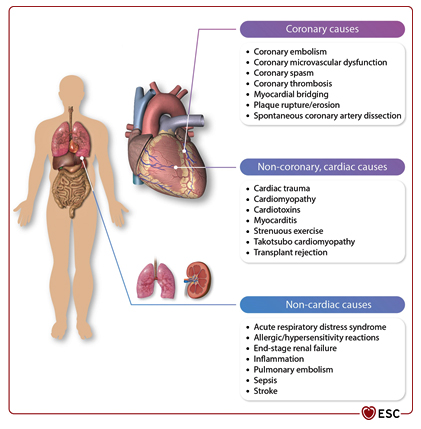
MINOCA can be considered a COMMON term that encompasses a heterogeneous group of underlying causes: coronary and non-coronary, which may include both cardiac and extracardiac disorders (Figure 1.).

Figure 1. MINOCA - a common term encompassing a heterogeneous group of coronary and non-coronary causes.
Source: https://academic.oup.com/view-large/figure/441045253/ehad191f15.tif

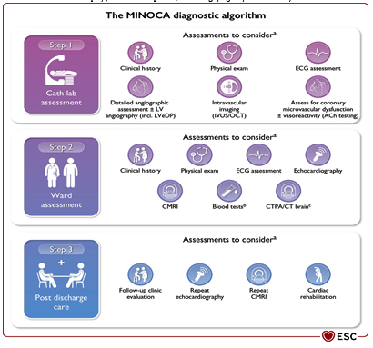
When the diagnosis is not clearly established after coronary angiography, MINOCA represents a working, rather than a final, diagnosis. It is crucial that clinicians conduct additional assessments and investigations to identify the underlying cause of MINOCA, which will enable a definitive diagnosis and appropriate treatment for the patient. Failure to identify the underlying cause of MINOCA may lead to inadequate or inappropriate therapy [1]. If the underlying cause of MINOCA cannot be determined solely through invasive coronary angiography (ICA), further assessments of microvascular function and coronary artery reactivity, as well as intravascular imaging, should follow. The term "functional coronary angiography" refers to the combination of standard coronary angiography with additional tests (e.g., testing for coronary microcirculation dysfunction and vasoreactivity) (Figure 2).

Figure 2. MINOCA diagnostic algorithm. Evaluation of patients with a working diagnosis of MINOCA.
Source: https://academic.oup.com/view-large/figure/441045256/ehad191f16.tif

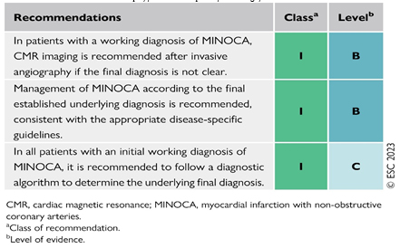
If the underlying cause of MINOCA cannot be determined through functional coronary angiography, non-invasive imaging is recommended (e.g., echocardiography, CMR, CT angiography, CT heart), according to the clinical indication. Cardiac magnetic resonance (CMR) is one of the key diagnostic tools for determining the underlying cause of MINOCA [7–11]. CMR can identify the underlying cause in up to 87% of patients with a working diagnosis of MINOCA and should be performed as soon as possible after patient admission, ideally during the initial hospitalization [12]. (Table 1)

Table 1. Recommendations for myocardial infarction with non-occlusive coronary arteries (MINOCA)
Source: https://academic.oup.com/view-large/441045259

Determining the underlying cause of MINOCA enables the initiation of appropriate therapy in accordance with the final diagnosis. Secondary preventive therapy should be considered for patients with evidence of coronary atherosclerotic disease and for the control of risk factors.
Treatment of Takotsubo syndrome is not based on prospective randomized controlled trials, so therapy is mainly based on supportive and empirical strategies [13,14]. Treatment of patients with myocarditis is carried out according to the recommendations of the working group for myocardial diseases of the European Society of Cardiology (ESC) [15,16].
Ischemia without infarction with non-occlusive coronary arteries (INOCA) has been described in the context of chronic coronary syndromes (CCS) [17,18].

CASE REPORT

A 60-year-old female patient presents with chest pain described as tightness and pressure at rest. She reports that the day before seeing the doctor, she felt chest pain resembling tightness and pressure. The pain occurs with exertion, most often when walking uphill, lasts longer than half an hour, radiates through the left side of the chest, and decreases with cessation of exertion, which corresponds to the clinical picture of unstable angina pectoris. She had no similar symptoms previously.
The family history was unremarkable. From the personal history, we learn that she is being treated for arterial hypertension and dyslipidemia and is a non-smoker.
Physical examination: Inspection revealed that the patient is conscious, oriented, mobile, of medium osteomuscular build and nutritional status, afebrile, eupneic, and with normal heart sounds. She appears to be moderately ill. The auscultatory findings on the heart and lungs are normal. Blood pressure is 160/90 mmHg. Heart rate is 75/min.
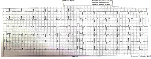
The electrocardiogram shows sinus rhythm, normal axis, a heart rate of 75/min, with ST segment depression in the anterolateral leads V4-V6 of 0.5 to 1 mm (Figure 3).

Figure 3. Electrocardiogram of the patient upon admission

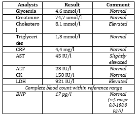
The patient was admitted with a diagnosis of acute coronary syndrome type unstable angina, and during hospitalization, the first cardiac biomarker, high-sensitivity troponin (hs-cTnI), was 4.3 ng/l, which was normal. The following day, hs-cTnI was repeated and found to be 79.2 ng/l, showing an increase of approximately 20 times from the first value, and 7 times higher than the upper reference limit (reference range 0.0-11.6 ng/l), which led to the definitive diagnosis of non-ST-elevation myocardial infarction.

Laboratory tests were performed:

Treated according to the NSTEMI protocol. The therapy included low-molecular-weight heparin, dual antiplatelet therapy (DAPT), the beta-blocker nebivolol, ACE inhibitor, calcium channel blocker, diuretic, proton pump inhibitor, and nitroglycerin as needed.
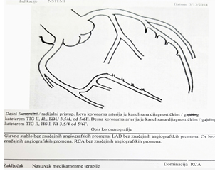
According to the NSTEMI criteria and the significant increase in serum troponin levels, coronary angiography was performed, which showed no significant angiographic changes in the main stem, LAD, Cx, and RCA (Figure 4)

Figure 4. Coronary angiography report

Given the absence of significant stenosis, medical and pharmacological therapy was continued. During the further course of hospitalization, to determine the cause of MINOCA, an echocardiogram was performed: concentric hypertrophy of the left ventricular walls, with normal systolic function, ejection fraction (EF) = 60%, without clear segmental abnormalities in contractility, which excludes a large infarction and supports the possibility of a subendocardial infarction in the presence of myocardial hypertrophy.
Throughout the course, the patient remained stable and cardially compensated. Further investigation of the definitive cause of non-occlusive infarction is recommended according to the MINOCA algorithm: stress echo test and cardiac magnetic resonance imaging (CMRI), and in the event of worsening symptoms, functional coronary angiography with intravascular ultrasound (IVUS) and optical coherence tomography (OCT), as well as microvascular dysfunction and coronary vasospasm tests [1].
At discharge, the patient was without chest pain, with a normal blood pressure value of 130/90 mmHg, and a normalized ECG: sinus rhythm, normal axis, heart rate 70/min, without ST-T changes (Figure 5)

Figure 5. ECG of the patient at discharge

Therapy at Discharge: DAPT, beta blocker nebivolol; ACE inhibitor, calcium channel blocker and diuretic as a fixed combination, proton pump inhibitor, and nitroglycerin as needed.

DISCUSSION

According to the fourth universal definition of myocardial infarction, there are several subtypes of myocardial infarction. There is a group of patients with MI without angiographically obstructive coronary artery disease (without stenosis of 50% or more in a large epicardial vessel), and the term myocardial infarction without coronary artery obstruction (MINOCA) has been adopted for this entity. Prevalence is estimated at 6-8% [4], but it is heterogeneous and varies from 1% to 14% of patients with ACS [1]. MINOCA is more common in women, as well as in patients presenting as NSTEMI [4]. The criteria for diagnosing MINOCA are:
1. Criteria for diagnosing acute myocardial infarction:
a) Positive cardiac biomarkers (preferably cardiac troponin);
b) Clinical evidence of myocardial infarction, including: ischemic symptoms, new or presumed new significant ST-T changes or new left bundle branch block (LBBB), development of pathological Q waves, evidence by imaging method of newly developed myocardial viability loss and impaired regional wall motion, identification of coronary thrombus on angiography or autopsy,
2. Absence of coronary artery obstruction,
3. Absence of a clear clinical cause for the AMI presentation [19, 20].
The causes of troponin elevation, as the most sensitive markers, can be divided into coronary and non-coronary causes. Coronary causes include: vasospastic angina, coronary microvascular dysfunction, rupture/erosion of an atherosclerotic plaque, spontaneous coronary thrombosis or embolism, and overlooked obstructive CAD. Non-coronary causes can be cardiac disorders: myocarditis, Takotsubo cardiomyopathy, other cardiomyopathies (dilated, hypertrophic), and non-cardiac disorders such as pulmonary embolism, renal insufficiency, and sepsis [21, 22]. An individualized approach to therapy is essential.
Our patient presented with new-onset chest pain and was observed to have shallow ST depressions on the ECG. She was hospitalized in the intensive care unit and treated according to the non-STEMI acute coronary syndrome protocol. Coronary angiography was performed, after which she was diagnosed with MINOCA. Throughout the course, she had no recurrent angina and no dynamic electrocardiographic changes. She was discharged with recommendations for hygiene-dietary regime measures and pharmacological therapy. Patient education was provided on the importance of maintaining an ideal body mass index, a diet rich in fruits and vegetables, the significance of daily moderate physical activity, and achieving target blood pressure and cholesterol levels.

CONCLUSION

Myocardial infarction with non-obstructive coronary artery disease (MINOCA) is a relatively new entity in cardiology and encompasses a heterogeneous group of patients who do not have significant coronary artery obstruction on angiography. The aim of this paper is to highlight MINOCA as a relatively new entity in cardiology, the importance of early diagnosis and timely treatment, as well as the application of primary and secondary prevention of cardiovascular diseases with modification of risk factors. The presented patient, based on the symptoms of angina and ST segment depression on the ECG, was initially diagnosed as a classic acute coronary syndrome without persistent ST elevation but with significantly elevated troponin levels, as a myocardial infarction with probable severe coronary stenosis or occlusion. However, the normal coronary angiography result classified her into the relatively rare MINOCA group. The patient was treated according to the acute coronary syndrome protocol, and during the course of hospitalization, there were no complications. She was discharged for further home treatment with pharmacological therapy, including dual antiplatelet therapy, and advice on hygiene-dietary measures to modify risk factors. The patient was advised to have regular check-ups and was referred for cardiac magnetic resonance imaging, which, according to the MINOCA algorithm, would clarify the cause of the infarction.

REFERENCE:

  1. Byrne RA, Rossello X, Coughlan JJ, Barbato E, Berry C. et al. 2023 ESC Guidelines for the management of acute coronary syndromes: Developed by the task force on the management of acute coronary syndromes of the European Society of Cardiology (ESC). European Heart Journal, 2023; 44 (38): 3720–3826. doi.org/10.1093/eurheartj/ehad191.
  2. Grech ED, Ramsdale DR. Acute coronary syndrome: unstable angina and non-ST segment elevation myocardial infarction. B M J 2003;326:259-1261. DOI: 10.1136/bmj.326.7401.1259.
  3. Miljuš D. Mickovski Katalina N. Bozic Z. Registar za akutni koronarni sindrom u Srbiji, 2022. Institut za javno zdravlje dr Milan Jovanović Batut. 2022. dostupno na: http://www.batut.org.rs/index.php
  4. Thygesen K, Alpert JS, Jaffe AS, Chaitman BR, Bax JJ, Morrow DA, et al. ESC Scientific Document Group. Fourth universal definition of myocardial infarction (2018) Eur Heart J. 2019;40:237–69. Doi: https://www.ahajournals.org/doi/10.1161/CIR.0000000000000617
  5. Hinić, S. Akutni koronarni sindrom bez perzistentne elevacije ST segmenta - šta su NAM donele nove preporuke. Galenika Medical Journal. 2022;1(1):105-110. DOI: https://scindeks-clanci.ceon.rs/data/pdf/2812-8575/2022/2812-85752201105H.pdf
  6. Čolaković G., Bogunović S., Anđelić S., Čolaković N. Zbrinjavanje pacijenata sa AKS u GZZHMP Beograd. Naučni časopis urgentne medicine - Halo 194, 2018;24(2):93-101.
  7. Eitel I, Behrendt F, Schindler K, Kivelitz D, Gutberlet M, Schuler G, et al. Differential diagnosis of suspected apical ballooning syndrome using contrast-enhanced magnetic resonance imaging. Eur Heart J. 2008;29:2651–2659. https://doi.org/10.1093/eurheartj/ehn433
  8. Eitel I, von Knobelsdorff-Brenkenhoff F, Bernhardt P, Carbone I, Muellerleile K, Aldrovandi A, et al. Clinical characteristics and cardiovascular magnetic resonance findings in stress (takotsubo) cardiomyopathy. JAMA 2011;306:277–286. https://doi.org/10.1001/jama.2011.992
  9. Ferreira VM, Schulz-Menger J, Holmvang G, Kramer CM, Carbone I, Sechtem U, et al. Cardiovascular magnetic resonance in nonischemic myocardial inflammation: expert recommendations. J Am Coll Cardiol 2018;72:3158–3176. https://doi.org/10.1016/j.jacc.2018.09.072
  10. Lurz P, Luecke C, Eitel I, Föhrenbach F, Frank C, Grothoff M, et al. Comprehensive cardiac magnetic resonance imaging in patients with suspected myocarditis: the MyoRacer-trial. J Am Coll Cardiol 2016;67:1800–1811. https://doi.org/10.1016/j.jacc.2016.02.013
  11. Reynolds HR, Maehara , Kwong RY, Sedlak T, Saw J, Smilowitz NR, et al. Coronary optical coherence tomography and cardiac magnetic resonance imaging to determine underlying causes of myocardial infarction with nonobstructive coronary arteries in women. Circulation 2021;143:624–640. https://doi.org/10.1161/circulationaha.120.052008
  12. Pathik B, Raman B, Mohd Amin NH, Mahadavan D, Rajendran S, McGavigan AD, et al. Troponin-positive chest pain with unobstructed coronary arteries: incremental diagnostic value of cardiovascular magnetic resonance imaging. Eur Heart J Cardiovasc Imaging 2016;17:1146–1152. https://doi.org/10.1093/ehjci/jev289
  13. Lyon AR, Bossone E, Schneider B, Sechtem U, Citro R, Underwood SR, et al. Current state of knowledge on Takotsubo syndrome: a Position Statement from the Taskforce on Takotsubo Syndrome of the Heart Failure Association of the European Society of Cardiology. Eur J Heart Fail 2016;18:8–27. https://doi.org/10.1002/ejhf.424
  14. Ghadri JR, Wittstein IS, Prasad A, Sharkey S, Dote K, Akashi YJ, et al. International expert consensus document on Takotsubo syndrome (Part II): diagnostic workup, outcome, and management. Eur Heart J 2018;39:2047–2062. https://doi.org/10.1093/eurheartj/ehy077
  15. Adler Y, Charron P, Imazio M, Badano L, Barón-Esquivias G, Bogaert J, et al. 2015 ESC Guidelines for the diagnosis and management of pericardial diseases: the Task Force for the diagnosis and management of pericardial diseases of the European Society of Cardiology (ESC) endorsed by: the European Association for Cardio-Thoracic Surgery (EACTS). Eur Heart J 2015;36:2921–2964. https://doi.org/10.1093/eurheartj/ehv318
  16. Caforio AL, Pankuweit S, Arbustini E, Basso C, Gimeno-Blanes J, Felix SB, et al. Current state of knowledge on aetiology, diagnosis, management, and therapy of myocarditis: a position statement of the European Society of Cardiology Working Group on Myocardial and Pericardial Diseases. Eur Heart J 2013;34:2636–2648,2648a-2648d. https://doi.org/10.1093/eurheartj/eht210
  17. Ford TJ, Stanley B, Good R, Rocchiccioli P, McEntegart M, Watkins S, et al. Stratified medical therapy using invasive coronary function testing in angina: the CorMicA trial. J Am Coll Cardiol 2018;72:2841–2855. https://doi.org/10.1016/j.jacc.2018.09.006
  18. Kunadian V, Chieffo A, Camici PG, Berry C, Escaned J, Maas AHEM, et al. An EAPCI expert consensus document on ischaemia with non-obstructive coronary arteries in collaboration with European Society of Cardiology Working Group on Coronary Pathophysiology & Microcirculation Endorsed by Coronary Vasomotor Disorders International Study Group. Eur Heart J. 2020;41:3504–3520. https://doi.org/10.1093/eurheartj/ehaa503.
  19. Agewall, S., Beltrame, J.F., Reynolds, H.R., et al. On behalf of the WG on Cardiovascular Pharmacotherapy: ESC working group position paper on myocardial infarction with non-obstructive coronary arteries. Eur Heart J, 2017;38(3):143-153. Doi: 10.1093/eurheartj/ehw149
  20. Emiš-Vandlik N, isar. MINOCA. NČ UM Halo 194. 2019; 25(3):165-172. DOI: https://doi.org/10.5937/Halo1903165E
  21. Agewall S, Giannitsis E, Jernberg T, Katus H. Troponin elevation in coronary vs. non-coronary disease. Eur Heart J 2011;32:404–411. DOI:10.1093/eurheartj/ehq456
  22. Parwani P, Kang N, Safaeipour M, Mamas M, Wei J, et al. Contemporary Diagnosis and Management of Patients with MINOCA. 2023;25(6):561-570. doi:10.1007/s11886-023-01874-x.


      [ Contents ] [ INDEX ] <<< ] >>> ]      


Timočki medicinski glasnik, Zdravstveni centar Zaječar
Journal of Regional section of Serbian medical association in Zajecar
Rasadnička bb, 19000 Zaječar, Srbija
E-mail: tmglasnik@gmail.com

Pretraživanje / Site Search

  www.tmg.org.rs


      Design: Infotrend  
         

counter on myspace