Journal of Regional Section of Serbian Medical Association in Zajecar

Year 2026     Vol 51     No 1
     
      [ Contents ] [ INDEX ] <<< ] >>> ]      
               
   
Review article

UPDATES IN THE 2025 ESC GUIDELINES FOR MYOCARDITIS AND PERICARDITIS: AN INTEGRATIVE APPROACH TO INFLAMMATORY MYOPERICARDIAL SYNDROMES AND IMPLICATIONS FOR CLINICAL PRACTICE

Dušan Bastać (1), Zoran Joksimović (1), Mila Bastać (2), Pavle Nešović (1)

(1) INTERNAL MEDICINE PRACTICE "DR BASTAĆ" ZAJEČAR; (2) MEDSCAN TADIĆ DIJAGNOSTIKA, ZAJEČAR


  Download in pdf format   Abstract: Introduction: Myocarditis is an inflammatory disease of the myocardium that can present with highly heterogeneous clinical manifestations, ranging from asymptomatic forms to fulminant heart failure and sudden cardiac death. The aim of this review paper is to present contemporary diagnostic and therapeutic approaches according to the latest ESC recommendations, integrating clinical experience and emphasizing the need for further research. LITERATURE REVIEW: In 2025, the European Society of Cardiology (ESC) published the first integrated guidelines addressing the diagnosis and treatment of myocarditis and pericarditis, introducing the new concept of inflammatory myopericardial syndrome (IMPS). This umbrella term recognizes the frequent clinical overlap between these two entities and their shared pathophysiological mechanisms. The paper analyzes key updates in classification, diagnostics, genetic evaluation, and therapeutic approaches, with particular emphasis on revised cardiac magnetic resonance (CMR) criteria (Lake Louise criteria), expanded indications for endomyocardial biopsy (EMB), and innovations in the treatment of pericarditis, including interleukin-1 inhibitors. The diagnostic paradigm for myocarditis has been changed. The discussion includes a comparison of ESC recommendations with American ACC/AHA guidelines, as well as contributions from domestic literature, particularly studies in the field of diastolic stress testing and inflammatory cardiomyopathies. The COVID-19 pandemic has further highlighted myocarditis as a potential complication of viral infections. CONCLUSION: The contemporary approach to myocarditis is shifting the paradigm by introducing the concept of inflammatory myopericardial syndrome and involves integrated diagnostics and therapy in accordance with the latest ESC guidelines, recognizing this syndrome as a clinically significant entity. Advances in the use of cardiac magnetic resonance imaging, broader indications for endomyocardial biopsy, and the introduction of targeted therapies, including interleukin-1 inhibitors, enable more precise diagnosis and individualized treatment strategies. Despite these advances, the heterogeneity of clinical presentation remains a challenge in everyday practice. Further research is necessary to improve understanding of the pathophysiology and to optimize the treatment of these patients..
Key words: Myocarditis, pericarditis, inflammatory myopericardial syndrome, ESC guidelines, cardiac magnetic resonance imaging, COVID-19, endomyocardial biopsy.
 
     
      INTRODUCTION

The 2025 ESC guidelines represent a turning point in the approach to inflammatory heart diseases, unifying myocarditis and pericarditis within a single framework [1]. This decision stems from an increasingly clear understanding that these two entities are functionally, anatomically, and pathophysiologically closely related, and that treating them separately often leads to fragmentation in diagnosis and therapy. The new concept of inflammatory myopericardial syndrome (IMPS) serves as an umbrella term encompassing a clinical continuum ranging from isolated myocarditis, through combined myopericarditis and perimyocarditis, to isolated pericarditis, including complex mixed forms, up to chronic inflammatory cardiomyopathy and constrictive pericarditis [2–5, 6–8]. Such an integrative approach aims to improve collaboration among specialists and guide future research.
The term inflammatory cardiomyopathy has also been introduced, referring to chronic myocarditis associated with cardiac dysfunction and ventricular remodeling with a hypokinetic phenotype, with or without dilation. The role of cardiac magnetic resonance (CMR) is now central in diagnostics [9–11]. In the domestic literature, among others, contributions by the authors of this review and their collaborators have advanced the understanding of diastolic dysfunction and echocardiographic parameters in inflammatory cardiomyopathies [12–14], while endomyocardial biopsy is recommended in high-risk cases [1, 15–21].

EPIDEMIOLOGY AND CLASSIFICATION

The guidelines report an incidence of pericarditis ranging from 3 to 32 cases per 100,000 inhabitants per year, while the incidence of myocarditis is between 6 and 8 cases per 100,000 inhabitants [1,15]. Higher rates have been observed in men and younger adults. A particular challenge is the fact that a large number of subclinical and mild cases, including those diagnosed within the context of MINOCA (myocardial infarction with non-obstructive coronary arteries), remain undiagnosed [6,9], which may lead to underestimation of the true incidence and prevalence of chronic forms of the disease.
IMPS is classified into the following clinical entities: isolated pericarditis, isolated myocarditis, myopericarditis (predominant pericarditis with mild myocardial involvement), perimyocarditis (predominant myocarditis with pericardial features), and chronic inflammatory cardiomyopathy (chronic myocarditis with structural remodeling, dysfunction, and an arrhythmogenic substr [1].

ETIOLOGY AND PATHOPHYSIOLOGY

The etiology of myocarditis and pericarditis is heterogeneous. In developed countries, viral infections predominate (enteroviruses, adenoviruses, parvovirus B19, human herpesvirus 6, influenza virus, hepatitis C virus) [5,16,17], whereas in endemic regions tuberculosis remains an important cause of pericarditis, particularly in predisposed individuals with HIV infection. Bacterial causes (diphtheria, borreliosis, staphylococcal infections) are less common. Autoimmune mechanisms may lead to inflammation in the context of systemic diseases (lupus, sarcoidosis, vasculitis), while toxic agents (anthracyclines, alcohol, cocaine) and drugs (checkpoint inhibitors) can also induce myocarditis. A 
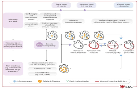
genetic basis plays an important role in susceptibility to viral infections and in determining the severity of the clinical presentation, with variants in sarcomeric and desmosomal genes being associated with myocarditis, and autoinflammatory diseases with recurrent pericarditis [6,8,15].
The pathophysiology of myocarditis is complex and involves acute, immune-mediated, and chronic phases of inflammation [5,6,17], progressing through several stages (Figure 1):
Acute phase: Direct myocardial injury caused by pathogen entry (e.g., viruses) into cardiomyocytes, leading to cell necrosis.
Immunological phase: Activation of the innate and adaptive immune response (macrophages, T-lymphocytes) with the release of cytokines and inflammatory mediators, resulting in further myocardial damage. Genetic variants in sarcomeric and desmosomal genes are increasingly being identified [1,18–20].
Chronic phase: In some patients, the inflammatory response persists, leading to progressive myocardial fibrosis, ventricular remodeling, and the development of dilated cardiomyopathy.

Figure 1. Stages of inflammatory myopericardial syndrome. AHA – anti-cardiac antibodies; AIDA – antibodies against the intercalated disc. Adapted from: Eur Heart J, Volume 46, Issue 40, 21 October 2025, Pages 3952–4041, https://doi.org/10.1093/eurheartj/ehaf192

CLINICAL PRESENTATION

The clinical presentation of IMPS is highly variable. According to the time course, myocarditis is classified into: acute (≤4 weeks), subacute (4–12 weeks), and chronic (>3 
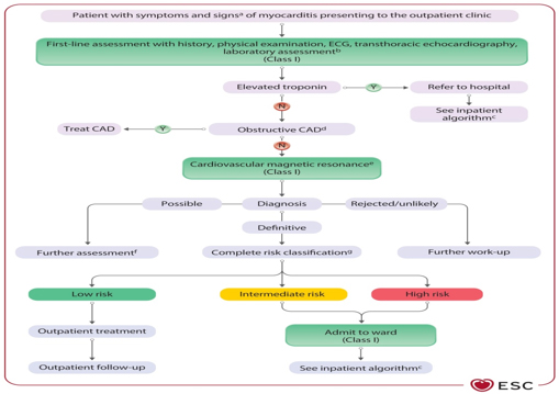
months, with persistent inflammation and remodeling).
Myocarditis may present with a wide spectrum of clinical manifestations, ranging from mild or asymptomatic forms to heterogeneous clinical phenotypes, including asymptomatic mild myocarditis, chest pain, perimyocarditis, heart failure, arrhythmic presentation, sudden cardiac death, and fulminant myocarditis with fulminant heart failure and cardiogenic shock [6,18–20].
Myocarditis is considered complicated when there is LVEF <50%, acute heart failure, ventricular arrhythmias, or high-degree atrioventricular (AV) block. Complicated forms may be associated with ventricular arrhythmias, AV blocks, and significantly reduced ejection fraction [7–9,18–20] (Figure 2).

Figure 2. Diagnostic algorithm and triage for outpatients with myocarditis. Adapted from: Eur Heart J, Volume 46, Issue 40, 21 October 2025, Pages 3952–4041, https://doi.org/10.1093/eurheartj/ehaf192

The term fulminant myocarditis [21] is reserved for patients presenting with cardiogenic shock and the most severe form of the disease, which often requires intensive treatment and mechanical circulatory support.
Pericarditis clinically manifests as dry (fibrinous), effusive, as an impending or acute cardiac tamponade, reversible partial constriction, or a chronic constrictive form [2,4]. Timely differentiation between inflammatory and non-inflammatory phenotypes is crucial.

DIAGNOSTIC APPROACH

The 2025 ESC recommendations significantly reshape the essence of the diagnostic pathway and disease staging, reflecting a paradigm shift in the diagnostic process. This is largely driven by the major role of cardiac magnetic resonance imaging (CMR), which has become the gold standard for diagnosing myocarditis. At the same time, the role of endomyocardial biopsy has been refined and is now mainly reserved for severe, unclear, or high-risk cases, as well as for guiding appropriate therapy based on pathological and histological characterization with immunohistochemistry and PCR detection of viral genomes in the
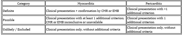
 myocardium.
Previously, diagnostic categories included: suspected, confirmed, and proven myocarditis. In the new framework, classification has been updated to: unlikely/excluded, possible, and definite myocarditis (Figure 3 and Tables 1 and 2).

Table 1. Diagnostic criteria and classification for inflammatory myopericardial syndrome (IMPS)
If diagnostic criteria for myocarditis and/or pericarditis are fulfilled

Table 2. Additional criteria (in addition to clinical presentation)

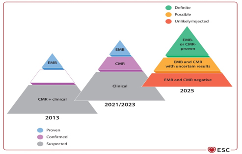
Figure 3. Paradigm shift in the clinical diagnosis of myocarditis
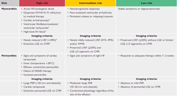
Basic diagnostic elements
Initial evaluation – biomarkers: Mandatory measurement of troponin (marker of myocardial injury/necrosis), high-sensitivity C-reactive protein (hsCRP, marker of inflammation), and natriuretic peptides BNP/NT-proBNP (markers of heart failure). Routine viral serologies are not recommended [1,3,5,19–20].
Electrocardiography (ECG): May show nonspecific ST-segment changes [5] (ST elevation/depression, T-wave changes, AV block, arrhythmias). There is no typical ECG pattern for myocarditis; however, ECG is rarely completely normal and is considered heterogeneous across different IMPS forms. Diffuse, concave upward ST-segment elevation is typical of pericarditis.
Echocardiography: A basic imaging modality for assessment of ventricular function (LVEF), presence of pericardial effusion, tamponade, and regional wall motion abnormalities. However, it often cannot definitively confirm inflammation [12–14].
If troponin is normal and LVEF is preserved, this indicates a favorable prognosis.
Non-invasive imaging – Cardiac Magnetic Resonance (CMR)
Cardiac magnetic resonance imaging is a key modality according to the revised Lake Louise criteria [1,10,11] and has become central in the diagnostic work-up of IMPS.
Diagnosis is established based on the revised Lake Louise criteria, which require at least one abnormality based on T1 mapping (indicating interstitial fibrosis or edema) and at least one abnormality based on T2 mapping (indicating myocardial edema) [11].
CMR enables detection of edema, necrosis, and fibrosis through late gadolinium enhancement (LGE), typically in an epicardial or mid-myocardial distribution, which helps differentiate inflammatory disease from myocardial ischemia and coronary artery disease.
The 2025 ESC recommendations emphasize an individualized approach based on clinical presentation and risk assessment (Table 3).1].

Table 3. Risk stratification algorithm and triage of patients with suspected myocarditis and pericarditis in outpatient settings. Hospitalization is recommended for all patients with myocarditis and high-risk pericarditis.

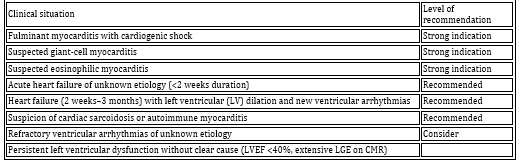
Invasive diagnostics – Endomyocardial biopsy (EMB)
Endomyocardial biopsy (EMB) is considered the gold standard in severe forms of disease [17,21,22], providing definitive diagnosis and etiological classification. Indications have been expanded compared with previous guidelines and are now reserved for high-risk cases (Table 4). Molecular biological (PCR) analysis for viral genomes is mandatory prior to initiation of immunosuppressive therapy [21–23].

TABLE 4. Indications for endomyocardial biopsy (EMB) according to ESC 2025 [21]

GENETIC TESTING

There is increasing evidence of an association between myocarditis and inherited cardiomyopathies. The ESC 2025 guidelines recommend genetic testing in selected patients with familial forms and recurrent pericarditis [1]. Studies highlight mutations in desmosomal genes as part of an inherited predisposition [1], particularly in patients with high diagnostic yield, such as those with:

  • Family history of cardiomyopathy or sudden cardiac death
  • Arrhythmic presentation of disease
  • Presence of septal or “ring-like” LGE on CMR
  • Persistent systolic dysfunction without recovery
  • Recurrent myocarditis or pericarditis (a genetic cause is identified in approximately 15% of patients with recurrent pericarditis) [1]

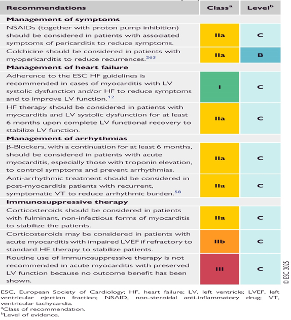
THERAPY OF MYOCARDITIS

Therapeutic management is individualized and depends on etiology, clinical presentation, and hemodynamic status [1,6] (Table 5). In stable patients, analgesics and NSAIDs are used, with the addition of colchicine when pericardial symptoms are present [1,31]. In heart failure, standard HF guideline-directed medical therapy is applied (ACE inhibitors/ARNI, beta-blockers, mineralocorticoid receptor antagonists, SGLT2 inhibitors) [6,29].
Immunosuppressive therapy is reserved for virus-negative forms [21] or specific entities such as giant-cell and eosinophilic myocarditis [22–24]. In fulminant myocarditis, mechanical circulatory support (VA-ECMO) is recommended [1,20].
The treatment of myocarditis is systematized into five key domains:

  1. General measures and symptomatic therapy
    Restriction of physical activity for 3–6 months. In the acute phase, in hemodynamically stable patients, analgesics, non-steroidal anti-inflammatory drugs (NSAIDs), and colchicine are used if pericardial symptoms are present.
  2. Heart failure therapy
    In patients with heart failure, standard guideline-directed therapy is used: ACE inhibitors or ARNI, beta-blockers, mineralocorticoid receptor antagonists (MRA), sodium-glucose cotransporter-2 inhibitors (SGLT2i), and diuretics in cases of congestion.
  3. Immunosuppressive therapy
    Used only in non-infectious etiologies and after exclusion of active viral infection in the myocardium by PCR analysis of EMB samples. Indications include:
    Virus-negative inflammatory (autoimmune) myocarditis
    Giant-cell myocarditis (strong recommendation)
    Eosinophilic myocarditis (strong recommendation)
    Myocarditis associated with systemic autoimmune diseases (e.g., sarcoidosis)
    Commonly used agents include corticosteroids (prednisone), azathioprine, mycophenolate mofetil, and cyclosporine [1].
  4. Antiarrhythmic therapy and devices
    Antiarrhythmic drugs are used for symptomatic arrhythmias. Implantable cardioverter-defibrillator (ICD) implantation is considered in patients with persistent LV dysfunction after at least 3 months of optimal therapy. A wearable defibrillator (vest-type device) may be used as a temporary option for 3–6 months [1].
  5. Mechanical circulatory support (MCS)
    In fulminant myocarditis with cardiogenic shock, MCS serves as a “bridge to recovery” or “bridge to transplantation.” Recommended modalities include intra-aortic balloon pump (IABP) and veno-arterial ECMO (VA-ECMO) as the most effective support strategies [20–21].].

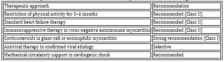
Table 5. Therapeutic recommendations for myocarditis according to ESC 2025

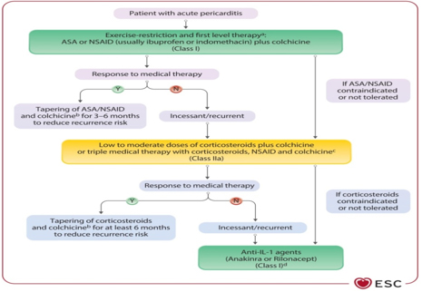
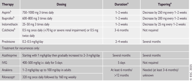
TREATMENT OF PERICARDITIS

First-line therapy: Aspirin or NSAIDs in combination with colchicine for at least 3 to 6 months (Class I A recommendation) [1–4].
Second-line therapy:

  • Corticosteroids are considered second-line treatment for refractory forms [2]. They are used only when first-line therapy fails or is contraindicated.
  • Refractory pericarditis:
    Interleukin-1 (IL-1) inhibitors represent a novel therapeutic option for treatment-refractory and/or recurrent pericarditis, particularly in patients with markedly elevated hsCRP levels (Class I A recommendation in highly inflammatory phenotypes) (Figure 4 and Table 6).

Figure 4. Proposed algorithm for pharmacological treatment of pericarditis in adults (excluding interventional procedures and pericardiectomy). Adapted from: Eur Heart J, Volume 46, Issue 40, 21 October 2025, Pages 3952–4041, https://doi.org/10.1093/eurheartj/ehaf192

TABLE 6. Therapeutic protocol for the treatment of pericarditis. Adapted from: Eur Heart J, Volume 46, Issue 40, 21 October 2025, Pages 3952–4041, https://doi.org/10.1093/eurheartj/ehaf192

TABLE 7. SUMMARY OF ESC RECOMMENDATIONS FOR THE TREATMENT OF INFLAMMATORY MYOPERICARDIAL SYNDROME (IMPS)

SPECIAL POPULATIONS

Post-COVID myocarditis: May occur as a result of direct viral infection, systemic inflammatory response, or immune dysregulation (25–26). Diagnosis is often established by CMR, and therapy is mainly supportive. Post-COVID myocarditis and post-vaccination forms have been analyzed in several studies [23–28]. Rare cases of post-vaccination myocarditis after mRNA vaccines have been reported, most commonly in young males within several days after the second dose. The clinical course is usually mild, and the prognosis is favorable. The benefits of vaccination far outweigh the risks [27–30].
Children: Similar diagnostic criteria are applied, but caution is required with corticosteroid use due to potential effects on growth.
Pregnancy: Colchicine may be used for recurrence prevention in pericarditis (Class IIb C recommendation).
Elderly patients: Dose adjustment of colchicine is necessary, along with monitoring of renal function.

PROGNOSIS AND FOLLOW-UP

Prognosis depends on initial clinical presentation and etiology. The most important predictor of adverse outcome is biventricular dysfunction. Most patients with mild disease achieve full recovery. In a minority of cases, progression to dilated cardiomyopathy and chronic heart failure may occur [14,15].
Follow-up after acute myocarditis is systematic over the first 6–24 months and includes: clinical assessment, ECG, Holter monitoring, biomarkers (troponin, CRP), echocardiography, exercise testing, and CMR in selected cases [1].
Prognosis is influenced by the degree of left ventricular dysfunction and the presence of fibrosis on CMR [6,10,11]. Diastolic dysfunction may persist even after clinical recovery [12–14].

DISCUSSION

The ESC 2025 guidelines represent a significant conceptual advance with the introduction of IMPS, but also raise several
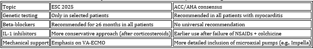
 controversies.
The most debated issues include: Central role of CMR: Although CMR has become indispensable, its position as a “new gold standard” carries the risk of overdiagnosis and limited accessibility.
Controversial beta-blocker recommendation [1,6]: The recommendation for beta-blocker use in all myocarditis patients for at least 6 months, regardless of EF, is based on limited evidence and is one of the most debated points in the guideline.
Genetic testing approach: Also controversial, being more limited in ESC guidelines compared with broader recommendations in ACC/AHA documents [3,4].
Insufficient commitment to clarifying the etiopathogenesis in the MINOCA population: The guidelines miss the opportunity to refine the definition of IMPS in patients with MINOCA, where myocarditis is one of the most common final findings.
Differences from the American recommendations (ACC/AHA) [1,3]: The European approach is more conservative regarding genetic testing (only selected patients vs. all), earlier use of IL-1 inhibitors in pericarditis (later compared to ACC) and does not include microaxial pumps (e.g. Impella) in the mechanical support algorithms, unlike the American guidelines.

Table 8. Comparison of ESC 2025 and ACC/AHA 2024 guidelines

The ESC 2025 guidelines introduce the IMPS concept, emphasize the role of CMR, and expand indications for EMB [1,10,17]. In the context of local clinical practice, the studies by the author of this literature review, Dr Dušan Bastać, significantly help bridge gaps in the practical application of diagnostic methods for 
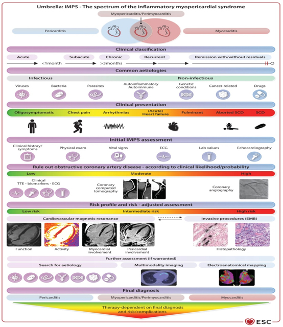
inflammatory myopericardial syndrome in Serbia [12–14].
His research on diastolic stress testing and echocardiographic markers indicates that diastolic dysfunction and elevated left ventricular filling pressures may persist even after recovery of systolic function (LVEF). This finding is consistent with the concept of inflammatory cardiomyopathy and the HFpEF phenotype (heart failure with preserved ejection fraction) in these patients.
These observations highlight the need for careful, multimodal follow-up even in patients with normalized ejection fraction.

Figure 5. Central illustration of the ESC guidelines on myocarditis and pericarditis. Adapted from: Eur Heart J, Volume 46, Issue 40, 21 October 2025, Pages 3952–4041, https://doi.org/10.1093/eurheartj/ehaf192

CONCLUSION

The ESC 2025 recommendations and guidelines represent the most comprehensive document to date, unifying myocarditis and pericarditis into a single concept—inflammatory myopericardial syndrome (IMPS), as illustrated in the figure (Figure 5: Central illustration of ESC guidelines on myocarditis and pericarditis).
The emphasis is on a multimodal diagnostic approach, with a central role of cardiac magnetic resonance imaging (CMR), rational use of endomyocardial biopsy (EMB), selective application of immunosuppressive therapy, and overall personalization of treatment strategies.
Despite significant progress, many recommendations are still based on expert consensus, highlighting the need for further high-quality research, particularly in areas such as optimal use of beta-blockers, novel antiviral therapies, genetic testing, and biological treatments.
The integration of domestic clinical experience shows that the guidelines are largely compatible with current practice, but also open new questions and research opportunities aimed at improving outcomes in patients with inflammatory heart diseases.

REFERENCE:

1. European Society of Cardiology. ESC Guidelines for the Management of Myocarditis and Pericarditis. Eur Heart J. 2025.
2. European Society of Cardiology. 2015 ESC Guidelines on Pericardial Diseases. Eur Heart J. 2015;36:2921–2964.
3. American College of Cardiology. ACC Expert Consensus Decision Pathway on Myocarditis. J Am Coll Cardiol. 2024.
4. American College of Cardiology. ACC Consensus on Pericardial Diseases. J Am Coll Cardiol. 2024.
5. Caforio ALP, Pankuweit S, Arbustini E, et al. Current state of knowledge on aetiology, diagnosis and therapy of myocarditis. Eur Heart J. 2022;43:399–421.
6. Ammirati E, Frigerio M, Adler ED, et al. Management of acute myocarditis and chronic inflammatory cardiomyopathy. Circulation. 2022;145:1722–1737.
7. Cooper LT. Myocarditis. N Engl J Med. 2009;360:1526–1538.
8. Tschöpe C, Ammirati E, Bozkurt B, et al. Myocarditis and inflammatory cardiomyopathy: current evidence and future directions. Nat Rev Cardiol. 2021;18:169–193.
9. Kindermann I, Barth C, Mahfoud F, et al. Update on myocarditis. J Am Coll Cardiol. 2022;79:1326–1342.
10. Friedrich MG, Sechtem U, Schulz Menger J, et al. Cardiovascular magnetic resonance in myocarditis: a JACC White Paper. J Am Coll Cardiol. 2009;53:1475–1487.
11. Ferreira VM, Schulz Menger J, Holmvang G, et al. CMR mapping techniques for myocarditis: consensus statement. JACC Cardiovasc Imaging. 2018;11:155 169.
12. Bastać D, Marković Z, Vasić B, et al. Dijastolni stres test u proceni bolesnika sa srčanom insuficijencijom. Zbornik radova Zdravstvenog centra Zaječar. 2017;1:45–49.
13. Bastać D, Vasić B, Nešović P, et al. Ehokardiografska procena dijastolne funkcije kod bolesnika sa miokarditisom. Timočki medicinski glasnik. 2018;43(2):73–78.
14. Bastać D, Vasić B, Marković Z, et al. Srčana insuficijencija sa očuvanom ejekcionom frakcijom – dijagnostički izazovi. Timočki medicinski glasnik. 2020;45(1):23–28.
15. Heymans S, Cooper LT, De Smet B, et al. Inflammatory cardiomyopathy. Eur Heart J. 2020;41:1358–1376.
16. Schultheiss HP, Kühl U, Cooper LT. Viral myocarditis. Nat Rev Cardiol. 2021;18:145–161.
17. Imazio M, Klingel K, Kindermann I, et al. Myocarditis management – current trends and future perspectives. Eur Heart J. 2022;43:1012–1027.
18. Kühl U, Pauschinger M, Noutsias M, et al. Viral persistence in myocardium is associated with progressive cardiac dysfunction. Circulation. 2005;112:1965–1970.
19. Ammirati E, Veronese G, Bottiroli M, et al. Contemporary management of myocarditis: diagnostic and therapeutic updates. Eur J Heart Fail. 2024;26:1–15.
20. Ammirati E, Cipriani M, Lilliu M, et al. Clinical presentation and outcome of myocarditis. Eur J Heart Fail. 2020;22:2117–2125.
21. McCarthy RE III, Boehmer JP, Hruban RH, et al. Long term outcome of fulminant myocarditis as compared with acute (nonfulminant) myocarditis. N Engl J Med. 2000;342:690–695.
22. Blauwet LA, Cooper LT. Myocarditis. Prog Cardiovasc Dis. 2010;52:274–288.
23. Frustaci A, Russo MA, Chimenti C. Randomized study on the efficacy of immunosuppressive therapy in virus negative inflammatory cardiomyopathy. N Engl J Med. 2009;360:1526–1538.
24. Cooper LT, Berry GJ, Shabetai R. Giant cell myocarditis. N Engl J Med. 1997;336:1860–1866.
25. Siripanthong B, Nazarian S, Muser D, et al. Recognizing COVID 19–related myocarditis: the possible pathophysiology and proposed diagnostic algorithm. Heart Rhythm. 2020;17:1463–1471.
26. Puntmann VO, Carerj L, Wieters I, et al. Outcomes of cardiovascular magnetic resonance imaging in patients recently recovered from COVID 19. JAMA Cardiol. 2020;5:1265–1273.
27. Mevorach D, Anis E, Cedar N, et al. Myocarditis after BNT162b2 mRNA COVID 19 vaccination. N Engl J Med. 2021;385:2140–2149.
28. Witberg G, Barda N, Hoss S, et al. Myocarditis after COVID 19 vaccination in patients under 40 years. N Engl J Med. 2021;385:1813–1822.
29. Bozkurt B, Kamat I, Hotez PJ. Myocarditis with COVID 19 mRNA vaccines. Circulation. 2021;144:471–484.
30. Oster ME, Shay DK, Su JR, et al. Myocarditis cases reported after mRNA based COVID 19 vaccination in the US. JAMA. 2022;327:331–340.


      [ Contents ] [ INDEX ] <<< ] >>> ]      


Timočki medicinski glasnik, Zdravstveni centar Zaječar
Journal of Regional section of Serbian medical association in Zajecar
Rasadnička bb, 19000 Zaječar, Srbija
E-mail: tmglasnik@gmail.com

Pretraživanje / Site Search

  www.tmg.org.rs


      Design: Infotrend  
         

counter on myspace