Journal of Regional Section of Serbian Medical Association in Zajecar

Year 2022     Vol 47     No 2-3
     
      [ Contents ] [ INDEX ] <<< ] >>> ]      
       

Page 119


Case report

Disconnection of arterial collateral as the cause of lower leg amputation after conquassation caused by petrol tiller. A case report

Ivan Golubović (1), Predrag Stojiljković (1), Mihailo Ille (2), Milan Radojković (3), Nemanja Jovanović (3), Milan Lazarević, Ivana Golubović (3), Ivan Milošević (2), Zoran Baščarević (4), Dejan Tabaković (5), Nebojša Mitić

(1)CLINIC FOR ORTHOPAEDIC SURGERY AND TRAUMATOLOGY, CLINICAL CENTER NIS, SERBIA; (2)CLINIC FOR ORTHOPAEDIC SURGERY AND TRAUMATOLOGY, CLINICAL CENTER BELGRADE, SERBIA; (3)FACULTY OF MEDICINE, UNIVERSITY OF NIS, SERBIA; (4)INSTITUTE OF ORTHOPAEDIC SURGERY “BANJICA”, BELGRADE, SERBIA; (5)CLINIC FOR ORTHOPAEDIC SURGERY AND TRAUMATOLOGY, CLINICAL CENTER KOSOVSKA MITROVICA, SERBIA


  Download in pdf format   ABSTRACT: Introduction. Leg conquassationconquassation caused by petrol tiller is one of the most severe injuries in bone and joint traumatology. Firm strokes by sharp tiller blades produce strong force that easily damages both soft tissues and bones. Since tillers are used in soil processing, the wounds are highly contaminated with dirt and fertilizers, hence the anaerobic spore-forming bacilli, such as tetanus and gas gangrene pathogens. Casereport. This paper presents the treatment of a 69 years old man with chronic arterial insufficiency of the lower extremities who suffered severe injury of the lower leg (IIIB open tibial fracture according to Gustillo) by petrol tiller while performing agricultural work. Due to the absence 
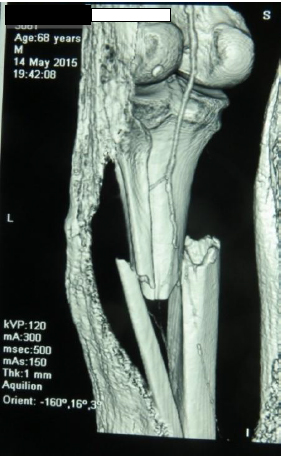
of pulsations, Multislice CT angiography and arteriography were performed. Arteriography of the injured leg confirmed chronic occlusion of the anterior tibial artery and numerous stenotic lesions of the peroneal and posterior tibial arteries. Posterior tibial artery was chronically occluded in its distal part and connected to the foot with moderately developed collateral arteries which provided the viability of the injured leg. Despite undertaken basic principles of treatment of this serious injury (primary surgical treatment of wounds, external fixation, reconstruction of soft tissue, antibiotic and anti-tetanus prophylaxis) due to infection and gangrene the treatment ended with lower leg amputation. Conclusion. Leg amputation can be expected in this type of injuries in cases of extensive destruction of tissue in the field of existing chronic arterial insufficiency in elderly patients, even in the absence of injury of main blood vessels due to traumatic disconnection of collateral in such patients.
Keywords: leg conquassation, IIIB open tibial fracture, external fixation, chronic arterial insufficiency, amputation of the lower leg


INTRODUCTION

Farmer is one of the most frequent professions in Serbia. Leg conquassation caused by engine tiller is among the most severe injuries in bone and joint traumatology. Firm strokes produced by sharp tiller blades produce strong force that easily damages both soft tissues and bones. Since tillers are used in soil processing, the wounds are highly contaminated with dirt and fertilizers, hence the anaerobic spore-forming bacilli, such as tetanus and gas gangrene pathogens. Skin and soft tissue destruction, comminution and bone defect, high level of both anaerobic and aerobic contamination and threatening infection make the treatment of these injuries, particularly open lower leg fracture, complex and challenging (1).
Meticulous irrigation of these wounds, removal of all foreign bodies and dirt and thorough surgical debridement of damaged tissue are crucial for successful prevention of infection, both non-specific and specific (tetanus and gas-gangrene). Also, fracture stabilization with external skeletal fixation, antibiotic therapy and anti-tetanus protection are mandatory. High-quality physical therapy following successful healing of soft tissue wounds and bone fractures is necessary for patient’s early recovery and faster return to everyday activities (2).
The most common complications of leg conquassation include soft tissue and bone infection, gas-gangrene, fracture malunion and finally amputation (3,4). Healing may be compromised and prognosis worsened by vascular insufficiency due to magistral vessels injury. Open Gustilo type IIIC fractures are among the most severe lower leg injuries.
These injuries often occur in older people over 60 who have co-morbidities. Chronic arterial insufficiency of lower extremity greatly complicates surgical treatment of this injury.
Aim of the study is to present a patient treated for severe lower leg and foot conquassation caused by petrol tiller during agricultural labor. We aimed to depict the specificities of such injury, the problems that may arise and the complications that may occur during the treatment of this severe trauma.

CASE REPORT

A 69-year old male patient was injured during soil processing by an engine tiller when the machine hit the hurdle in the ground, changed the direction and caused him severe both right and left lower legs and feet trauma with its sharp blades. Injuries included open left lower leg fracture Gustilo type IIIB with soft tissue defect, severe laceration of the left foot dorsum also with soft tissue defect and right lower leg laceration. He was initially admitted to the regional hospital emergency unit where his injuries were assessed and left leg plaster immobilization was done. Subsequently, the patient was referred to Orthopaedics and Traumatology Clinic, Clinical Center Nis where resuscitation and preoperative preparation were immediately performed. On examination, there was large skin and subcutaneous tissue defect on the front left lower leg with lacerated and ruptured tibialis anterior muscle tendon. X-rays revealed comminuted fracture of the left lower leg proximal third and left medial malleolus fracture. Left lower leg was deformed in the proximal third with complete functional impairment. There were crepitations during the movements and palpation of the fracture site. Both anterior and posterior tibial pulses were absent.
Multislice CT angiography revealed multiple stenoses in popliteal and tibioperoneal arteries of the injured leg, and also significant lesions in crural arteries (Figure 1). After preoperative preparation, surgery is done 9 hours after the injury, along with primary treatment of the wounds, fracture reduction and external fixation (Figure 2). In the absence of a pronounced bleeding, as well as violations of main blood vessels, there was no need for vascular reconstruction. Postoperatively angiography confirmed chronic occlusion of the anterior tibial artery and numerous stenotic lesions of the peroneal and posterior tibial arteries. Posterior tibial artery was chronically occluded in its distal part and connected to the foot with moderately developed collateral arteries which provided the viability of the injured leg (Figure 3). Also, additional multiple multilevel stenoses and occlusions were detected in the proximal arterial segments of our patient.

Figure1. Multislice CT angiography of injured lower leg

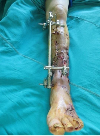
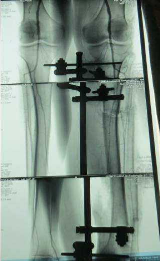
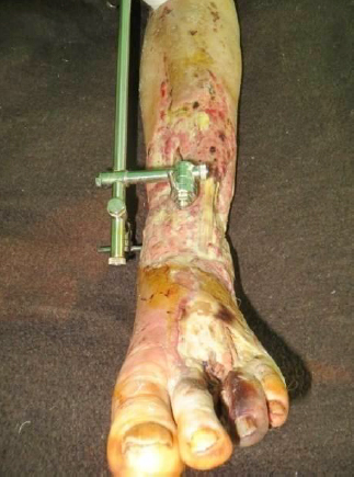
Figure 2. Left lower leg after the primary wound care and external skeletal fixation.


Figure 3. Postoperatively angiography of the lower leg.

The patient was administered anti-tetanus protection and anticoagulant prophylaxis of deep vein thrombosis and pulmonary thromboembolism (nadroparin 0,6mL/24hr). The patient received postoperative intravenous antibiotic therapy (ceftriaxone 2gr daily, amikacin 500mg/12hr and metronidazole 500mg/8hr). Vascular surgeon administered the medication therapy for chronic arterial 
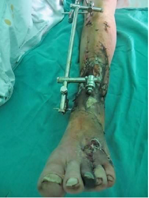
insufficiency of the lower extremities.
Subsequently, front lower leg and foot dorsum soft tissue necrosis occurred with the dry gangrene of the third toe (Figure 4).

Figure 4. Front left lower leg and foot dorsum soft tissue necrosis with the dry gangrene of the third finger.

Secondary wound debridement including necrosectomy and gangrenous third finger of the left foot amputation was performed in spinal anesthesia (Figure 5).

Figure 5. Left lower leg and foot after secondary wound debridement including necrosectomy and gangrenous third finger amputation

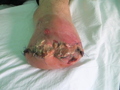
It was followed by general aggravation, severe wound infection, infection around the skeletal fixator nails and critical lower leg ischemia. A multidisciplinary team of orthopedist, vascular and plastic surgeons made a decision to amputate the lower leg for vital indication (Figure 6).

Figure 6. Severe infection of the amputation stump.

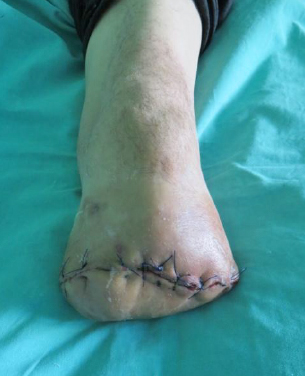
Perioperatively, the patient was administered 1750mL whole blood transfusion and 1500mL fresh frozen plasma. Postoperatively it was continued with antibiotics (ceftriaxone 2gr daily, clindamycin 600mg/12hr and vancomycin 1gr/12hr) and subcutaneous anticoagulant (nadroparin 0,6mL/24hr). Meticulous everyday wound cleaning and dressing was performed. However, infection and necrosis of the amputation stump developed. All the sutures were removed, debridement of the stump was done and it was left wide open. Thorough everyday wound care was continued. Seven days after the amputation secondary debridement of the stump and wound closure were performed (Figure 7). Postoperative course was uneventful. The stump healed and the sutures were removed. The patient was referred to physical therapy and limb prosthesis specialist.

Figure 7. Amputation stump after repeated debridement and secondary wound closure

DISCUSSION

Agriculture is one of the most important economy braches in Serbia. Limb injuries caused by petrol tiller almost always include sever skin and soft tissue destruction, magistral blood vessels injury, severe comminuted fracture and often traumatic amputation. These injuries are highly mutilating and may lead to death. Possibilities of tissue reconstruction are small and require multidisciplinary approach that includes orthopedist, vascular and plastic surgeons.
Lower leg conquassation caused by petrol tiller requires urgent surgical treatment providing patient’s satisfactory general condition. The management of the open lower leg fracture contaminated with soil includes meticulous wound irrigation, removal of all foreign bodies and dirt, thorough surgical debridement of damaged tissue, fracture stabilization with external skeletal fixation, antibiotic therapy, anti-tetanus protection and delayed wound closure (5).
Primary surgical care – debridement of the open fracture wound is crucial for prevention or successful treatment of infection. If possible, it has to be done within six hours after injury in order to prevent progressive wound contamination and infection, including gas gangrene, tetanus and osteitis. Wound smear, microbiological examination for contaminating microorganisms’ identification and their sensitivity to antibiotics (biogram and antibiogram) are necessary before primary treatment. First step is meticulous wound irrigation using saline and hydrogen peroxide (sometimes more than 10L) followed by detailed cleaning and removal of all foreign bodies – dirt, pieces of clothing and cellular debris. Debridement must include extensive surgical removal of devitalized soft tissue (skin, fat, fascia, muscle and bone) (6). Since necrotic muscle tissue represents the environment susceptible for both aerobic and anaerobic bacteria, special care has to be made during muscle debridement regarding adequate assessment of its color, consistence, contractility and bleeding. The surgical removal of a muscle tissue that does not bleed and tightens when touched, does not have natural roseate healthy color and does not look vital is mandatory. If necessary, open fracture wound debridement may be repeated after 24 or 48 hours (secondary debridement) after demarcation and exposure of further (new) tissue devitalization. Adequate primary surgical care is most important for the successful prevention of deep osteitis and leg salvation (7,8).
Further treatment includes bones reposition and external skeletal fixation which is the method of choice for lower leg open fracture stabilization except for Gustilo type I fractures when internal fixation is possible. External skeletal fixation provides optimal biomechanical conditions for successful fracture healing, good approach for wound care and does not interrupt knee and ankle joint movements. Postoperatively, patients are being mobilized early, start with knee and ankle movements and walking (9).
Problems related to external skeletal fixation include common soft tissue and bone infection around the device nails, especially if applied for more than six months. Edwards and al. reported 50 (29,24%) patients with soft tissue infection and 4 (2,33%) with local osteitis around the nails in a study of 171 patients with open fracture treated with external skeletal fixation (10). Marsh et al. reported the incidence of 39 (38,61%) patients with complications related to device nails among 101 patients with open tibia fracture treated with external skeletal fixation and 10 of them required device replacement. However, in the same study low incidence of deep bone infection around fracture (6%) was observed (11).
Early aggressive soft tissue reconstruction during the first 7 days after the injury, in order to cover the fractured bone segments in patients with grade III open fractures, significantly reduces the risk of infection, fracture malunion/nonunion and amputation (12). Delayed wound closure is preferable and is performed after infection is definitely ruled out, using suturing or plastic and reconstructive surgery procedures (fasciocutaneous or microvascular flap), depending on the soft tissue defect size (1).
Early intravenous antibiotic therapy in patients with lower leg open fractures should be initiated immediately on admission. 3 antibiotics are usually administered for a period of 5 days to cover the entire bacterial flora as there is a massive contamination of the soil. After completed microbiological examination, further antimicrobial treatment should be administered according to antibiogram results and continued for additional 48-72 hours for types I and II open fractures and 120 hours for type III (6). Anti-tetanus protection is mandatory for all patients with open fractures.
Treatment outcome in such patient depends, among other factors, on the residual perfusion of the injured leg which can be diminished due to both injury of the magistral vessel and pre-existent chronic arterial insufficiency. Since our patient had marked advanced asymptomatic occlusive arteriosclerosis of the leg confirmed angiographically, the absence of clinically significant critical ischemia could only be explained with the functional collateral circulation that compensated serious perfusion deficit. Extreme trauma such as presented in our patient including the injury and exclusion of collateral arterial blood supply may lead to the critical leg ischemia and gangrene. The experience with our patient developing posttraumatic limb gangrene and amputation demonstrates the severity of the consequences of collateral circulation damage which is often inevitable in such injuries. Considering that the leg viability in patients with chronic arterial insufficiency may depend on the patency of no more than one sole seemingly insignificant collateral vessel 1-2mm in diameter, its damage due to trauma or surgical ligation may reduce the perfusion to the critical ischemia and gangrene.
Amputation of the lower leg on account of contaminated environment requires delayed closure of the stump when there are no signs of infection.

CONCLUSION

Leg amputation, can be expected in conquassant lower leg injuries in cases of extensive destruction of tissue in the field of existing chronic arterial insufficiency in elderly patients, even in the absence of injury of main blood vessels due to traumatic disconnection of collateral.
Acknowledgement: This paper is part of the project III 41017 Virtual human osteoarticular system and its application in preclinical and clinical practice, funded by the ministry of Education and Science of Republic of Serbia.

LITERATURE:

  1. Golubović Z, Stojiljković P, Mačukanović-Golubović L, Milić D, Milenković S et al. Lečenje otvorenih preloma potkolenice metodom spoljne skletne fiksacije. Vojnosanitet Pregl. 2008; 65(5): 343-7.
  2. Cross WW, Swiontkowski MF. Treatment principles in the management of open fractures. Indian J Orthop. 2008; 42(4): 377-86.
  3. Kohlprath R, Assal M, Uc¸kay I, Holzer N, Hoffmeier P, Suva D. Open fractures of the tibia in the adult: surgical treatment and complication. Rev Med Suisse 2011; 7(322): 2482–4.
  4. Gustilo RB, Mendoza RM, Williams DN. Problems in the management of type III (severe) open fractures. A new classification of type III open fractures. J Trauma. 1984; 24: 742-6.
  5. Golubovic I, Stojiljkovic P, Golubovic Z, Jeremic S, Radojkovic M, Stevanovic G et al. Leg conquassation caused by petrol tiller with open lower leg fracture. Acta Medica Medianae 2014; 53(1): 34-41.
  6. Golubovic I, Ristic B, Stojiljkovic P, Ciric M, Golubovic I, Radovanovic Z et al. Results of open tibial fracture treatment using external fixation. Srp Arh Celok Lek. 2016; 144(5-6): 293-9.
  7. Golubovic I, Vukasinovic Z, Stojiljkovic P, Golubovic Z, Stamenic S, Najman S. Lecenje otvorenih segmentnih preloma potkolenice metodom spoljne skeletne fiksacije. Srp Arh Celok Lek. 2012; 140(11-12): 737-7.
  8. Kindsfater K, Jonassen EA. Osteomyelitis in grade II and III open tibia fractures with late debridement. J Orthop Trauma 1995; 9(2): 121−7.
  9. Mitkovic M, Bumbasirevic M, Golubovic Z, Micic I, Mladenovic D, Milenkovic S, et al. New concept in external fixation. Acta Chir Iugosl 2005; 52(2): 107−11.
  10. Edwards CC, Simmons SC, Browner BD, Weigel MC. Severe open tibial fractures. Results treating 202 injuries with external fixation. Clin Orthop Relat Res. 1988; 230: 98- 115.
  11. Marsh JL, Nepola JV, Wuest TK, Osteen D, Cox K, Oppenheim W. Unilateral external fixation until healing with the dynamic axial fixator for sever open tibial fractures. J Orthop Trauma. 1991; 5: 341-8.
  12. Caudle RJ, Stern PJ. Severe open fractures of the tibia. J Bone Joint Surg Am 1987; 69(6): 801−7.


      [ Contents ] [ INDEX ] <<< ] >>> ]      


Timočki medicinski glasnik, Zdravstveni centar Zaječar
Journal of Regional section of Serbian medical association in Zajecar
Rasadnička bb, 19000 Zaječar, Srbija
E-mail: tmglasnik@gmail.com

Pretraživanje / Site Search

  www.tmg.org.rs


      Design: Infotrend  
         

counter on myspace